Skull Anatomy: Facial Bone Mnemonic
Prefer a Video? Sit Back, Relax, and Enjoy!!
Save yourself time and studying with the above video full of animations, visuals, and tricks to remember everything discussed below!
Don’t miss out on the other EZmed videos people are using to make medicine easy! Click below to check them out, and join to save time and help you study!
Introduction
The skull is made up of 22 bones that articulate with each other - 8 cranial bones and 14 facial bones.
The remaining 7 bones in the head (6 auditory ossicles and 1 hyoid bone) do not articulate with the rest of the skull, and they are often referred to as accessory bones of the skull as a result.
In this post, we will discuss the facial bones along with their anatomy and landmarks using labeled diagrams.
By definition, the facial bones form the face and are the osseous structures around the mouth, nasal cavity, and orbits.
Alternatively, the cranial bones form the surrounding cranium that encloses and protects the cerebrum, cerebellum, and brainstem.
Every EZmed post is filled with simple tricks to learn the material, and today we will use an easy mnemonic to remember the facial bone names and anatomy.
Make sure to also check out the EZmed cranial bone mnemonic which will help you remember the names and anatomy of the cranial bones as well!
Let’s get started!
Image: The skull is made up of 8 cranial bones and 14 facial bones, for a total of 22 bones (excluding the ear ossicles and hyoid). We will focus on the 14 facial bones in this post.
Facial Bone Mnemonic
The skull has 14 facial bones as mentioned above.
Facial bone anatomy is important to understand, especially when managing an injury to the face.
For example, the Le Fort system is used to classify various facial fractures based on the location of injury and the bones involved.
The names of the facial bones can be remembered using the following mnemonic:
“My Mandible Chews Nine Very Large Zucchini Pizzas”
My = Maxillary (2)
Mandible = Mandible
Chews = Conchae (2)
Nine = Nasal (2)
Very = Vomer
Large = Lacrimal (2)
Zucchini = Zygomatic (2)
Pizzas = Palatine (2)
The (2) denotes a pair, or 2, of those bones in the skull.
We are now going to discuss the anatomy and important features of each facial bone in the order of the mnemonic.
Image: The above mnemonic will help you remember the names of the facial bones.
Maxillary Bones
The maxillary bones are shown in purple below.
There are 2 maxillary bones and they are fused together.
They form part of the front of the face, the nasal passageway, the hard palate, and the inferior portion of the orbits.
There are a couple features to know about the maxillary bones: the infraorbital foramen and the incisive foramen.
Foramen, or foramina (plural), is a passage or hole in the bone.
First, there is a foramen located under each orbit, known as the infraorbital foramen.
The infraorbital foramen allows for the passage of the infraorbital nerve, artery, and vein.
Nerve blocks can be performed at the infraorbital foramen (approached from inside the mouth) to anesthetize the lower eyelid, side of the nose, upper lip, and upper teeth including the incisors, canines, premolars, and root of the first molar.
Next, there is another foramen involving the maxillary bones called the incisive foramen, also known as the anterior palatine foramen.
The incisive foramen is located at the roof of the mouth behind the incisors, as the name suggests.
It is an opening in the hard palate that allows for the passage of the sphenopalatine artery and nasopalatine nerves.
Lastly, the maxillary sinuses are located within the maxillary bones. There will be a future EZmed post that will go into much more detail about the sinuses.
Mandible
As we continue through the mnemonic, the next bone is the mandible (jaw) shown in turquoise/green below.
The mandible articulates with the temporal bone, which is one of the cranial bones.
The mandible has several important features to know.
First, there are 2 foramina located on the outer mandible (one on each side) called the mental foramina. They serve as a passageway for the terminal branches of the inferior alveolar nerve and blood vessels.
The mental foramen is another location point where nerve blocks can be performed to anesthetize the lower teeth and skin of the jaw and lower lip.
The next feature is the coronoid process, a superior anterior projection off the ramus. It serves as an attachment point for the temporal muscle.
Next, the condylar process is a superior posterior projection off the ramus. It serves as an articulation point between the mandible and temporal bone, ultimately allowing the jaw to open and close.
Lastly, there are 2 foramina located inside the mandible (one on each side) called the mandibular foramina. They serve as a passageway for the inferior alveolar nerve, artery, and vein.
The mandibular foramen is yet another location point where a nerve block can be performed to anesthetize the inferior alveolar nerve and its innervation to the lower teeth.
Conchae
The next pair of facial bones are the nasal conchae, specifically the inferior nasal conchae shown in red/maroon below.
They are best seen in a sagittal view of the nose, as they are located within the nasal cavity.
There are 3 pairs of nasal conchae: superior, middle, and inferior.
The superior and middle conchae are formed from the ethmoid bone, while the inferior nasal conchae are their own separate facial bones.
The nasal conchae serve to allow for rapid warming and humidification of air.
Nasal
As we move through the mnemonic, the nasal bones are next shown in beige/tan below.
There are no significant features to know about the nasal bones.
Just remember that most of the nose is made up of cartilage, and the only bony part of the nose is the bridge of the nose where the nasal bones are located.
Vomer
Next is the vomer bone shown in pink below.
It is located in the midline of the nasal cavity and can best be seen in a sagittal view of the nose.
The vomer bone forms the inferior portion of the nasal septum, which is what divides the right and left nares/nasal airway.
The vomer bone articulates with the sphenoid bone, ethmoid bone, maxillary bones, and palatine bones.
Lacrimal Bones
The next pair of facial bones are the lacrimal bones.
They are shown in pink in one of the images below and highlighted in yellow in the other.
The lacrimal bones are the smallest bones in the skull and make up part of the medial aspect of the each orbit.
The word “lacrima” means tears in latin, and this will help you remember the function of the lacrimal bones.
The shape of each lacrimal bone helps form the canals for the lacrimal apparatus which is involved in tear production and drainage.
Zygomatic Bones
The next pair of facial bones are the zygomatic bones shown in orange below.
The zygomatic bones form part of each orbit as well as the cheekbone that can be felt on either side of the face.
One of the features of the zygomatic bone is the temporal process, a posterior lateral projection.
The temporal process of the zygomatic bone joins with the temporal bone, specifically the zygomatic process of the temporal bone.
Together, the temporal process of the zygomatic bone and the zygomatic process of the temporal bone join to form the zygomatic arch, which is the cheekbone felt on either side of the face.
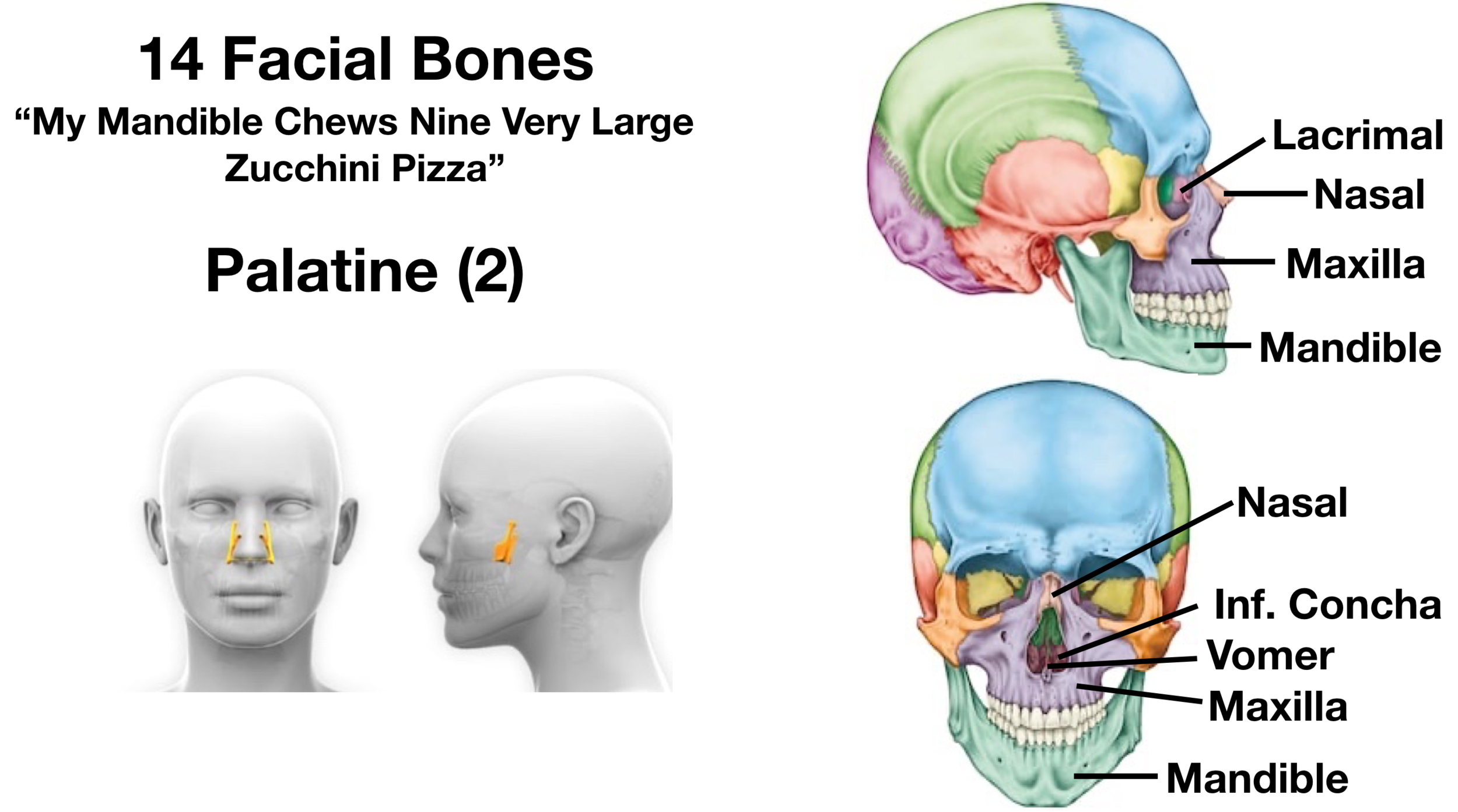
Palatine Bones
The final pair of facial bones are the palatine bones.
They are deeper in the skull and are shown below in blue.
The palatine bones help form 3 different cavities including the orbits, nasal cavity, and oral cavity.
Specifically, they help form part of the floor of the orbits, the lateral walls and floor of the nasal cavity, and the roof of the oral cavity.
The palatine bones help form the posterior aspect of the roof of the mouth, while the maxillary bones help form the anterior aspect of the roof of the mouth.