Body Cavities and Membranes: Labeled Diagram, Definitions
Body cavities along with their organs and membranes simplified! Labeled diagrams, definitions, and lateral views included! High-yield flow chart and table of the dorsal, ventral, cranial, spinal, thoracic, pleural, pericardial, abdominal, and pelvic cavities!
Save Time with a Video!
Save time by watching the video first, then supplement it with the lecture below!
Click below to view the EZmed video library. Subscribe to stay in the loop!
Body Cavities and Membranes
Welcome to a high-yield review of the major body cavities!
We will discuss the definitions and features of the following body cavities:
Dorsal Cavity
Ventral Cavity
Cranial Cavity
Spinal Cavity
Thoracic Cavity
Pleural Cavities
Pericardial Cavity
Abdominopelvic Cavity
Abdominal Cavity
Pelvic Cavity
You will learn the main organs housed in each cavity, along with the membranes that line the cavity and the type of fluid present.
Labeled diagrams, lateral views, and concise explanations included!
By the end of the lecture you will know the entire flow chart and table below!
Let’s get right into it!
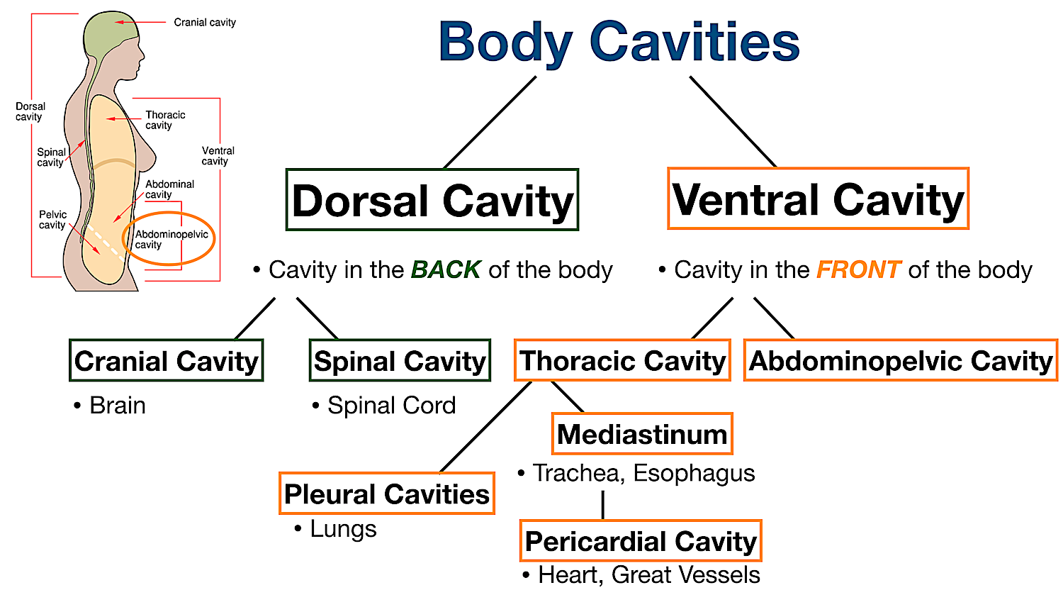
Body Cavities Labeled Diagram: Anatomy flow chart of the major body cavities and the main organs they house.
Body Cavities Labeled Diagram: Anatomy table of the major body cavities along with their organs, membranes, and fluid.
Body Cavity Definition
Before we review the main body cavities, let’s first define a body cavity.
A body cavity is a space or compartment in the body that houses organs or structures.
In other words, a body cavity is the space left over when internal organs are removed.
Body Cavity Definition: A space or compartment in the body that houses organs and structures.
Dorsal Cavity and Ventral Cavity
There are a number of different cavities in the body.
To make them easier to understand, let’s create a flow chart.
First, there are 2 main cavities in the body.
They include the dorsal cavity and the ventral cavity.
Body Cavities Labeled Diagram: There are 2 main cavities in the body, the dorsal cavity and the ventral cavity.
We know from the anatomical directional terms lecture that anterior means front or toward the front of the body, and posterior means back or toward the back of the body.
We also learned from the lecture another name for anterior is ventral, and another name for posterior is dorsal.
You can think of a ventriloquist for ventral, which literally translates to “stomach talker”.
If you point to your stomach then you are pointing to the front of your body, and this can help you remember ventral means front.
For dorsal, you can think of the dorsal fin on the back of a fish to help you remember dorsal means back.
Ventral (Anterior) and Dorsal (Posterior) Definitions: Ventral means front or toward the front of the body, and dorsal means back or toward the back of the body.
We also learned in the medical prefix lecture that “ventri-” means stomach, abdomen, toward the front, or the anterior aspect of the body.
We also discussed the prefix “dorso-” means back or posterior.
Ventral and Dorsal Medical Prefixes: “Ventri-” means anterior and “Dorsi-” means posterior.
Dorsal Cavity
Now that we know dorsal means back/posterior and ventral means front/anterior, we can apply those terms to the dorsal and ventral cavities.
Let’s start with the dorsal cavity.
We know dorsal means back or posterior, so the dorsal cavity is the cavity located in the back of the body.
The dorsal cavity is highlighted in red and labeled with a star below.
You can see how the dorsal cavity is situated behind or posterior to the ventral cavity.
The dorsal cavity houses the contents of the central nervous system including the brain and spinal cord - more on that soon!
Body Cavities Labeled Diagram: The dorsal cavity is located in the back of the body (red/stars) and houses the central nervous system including the brain and spinal cord.
Ventral Cavity
The ventral cavity is the cavity located in the front of the body, which makes sense because ventral means front or anterior.
The ventral cavity is highlighted in red and labeled with a star below.
You can see how the ventral cavity is situated in front of or anterior to the dorsal cavity.
The ventral cavity houses the contents within the chest, abdomen, and pelvis.
Body Cavities Labeled Diagram: The ventral cavity is located in the front of the body (red/star) and houses the organs/structures of the chest, abdomen, and pelvis.
Cranial Cavity
Now that we have a good understanding of the dorsal and ventral cavities, each cavity can be broken down even more.
The dorsal cavity can be subdivided into 2 main parts:
Cranial Cavity
Spinal Cavity
We will first discuss the cranial cavity followed by the spinal cavity.
Body Cavities Labeled Diagram: The dorsal cavity can be subdivided into the cranial cavity and spinal (vertebral) cavity.
Cranial Cavity Features
Location: Superior Dorsal Cavity
Enclosed By: Cranium/Skull
Houses: Brain
Fluid: Cerebrospinal Fluid
Membrane: Meninges
The cranial cavity is the superior portion of the dorsal cavity, as we can see highlighted in red and labeled by the star below.
The cranial cavity is enclosed by the cranium or skull, and it houses the brain.
The cranial cavity is filled with fluid called cerebrospinal fluid that helps protect and cushion the brain.
Each cavity is also lined with thin sheets of tissue called membranes.
The cranial cavity is lined by a 3 layer membrane called the meninges.
The meninges also help protect and cover the brain.
The 3 meningeal layers are the dura mater, arachnoid, and pia mater.
Cranial Body Cavity: Labeled diagram of the cranial cavity (red) along with its features, such as its organs (brain), membranes (meninges), and fluid (cerebrospinal fluid).
Cranial Cavity Anatomy
Let’s look closer at the cranial cavity and all of its features.
We are looking at a side view of the brain and skull below called a sagittal view.
As mentioned before, the cranial cavity is enclosed by the cranium or skull which is highlighted in green.
You can see how the skull has formed an empty space or cavity.
This space created by the skull is called the cranial cavity which is shown in yellow.
Cranial Body Cavity: Labeled diagram of the cranial cavity (yellow) which is enclosed by the cranium or skull (green).
We know the cranial cavity houses the brain which is colored in red below.
As mentioned above, the cranial cavity also contains fluid called cerebrospinal fluid or CSF.
The CSF helps protect and cushion the brain, along with other functions, and it is located in the subarachnoid space between the meningeal layers.
Remember we said the cranial cavity is lined by a 3 layer membrane called the meninges.
The pia mater is the innermost layer and closely adheres to the brain. The pia mater is the inner black line on the image, between the brain and CSF.
The dura mater is the outermost layer and the arachnoid is the middle layer. They are represented by the outer black line on the image, between the cranium and CSF.
The CSF is located in the subarachnoid space between the arachnoid and pia mater.
Cranial Body Cavity: Labeled diagram of the cranial cavity which houses the brain (red), and contains cerebrospinal fluid in the subarachnoid space (yellow) between the arachnoid and pia mater meningeal layers.
Let’s take a closer look at the layers associated with the cranial cavity.
Use the colored diagram in the bottom left of the image as a reference.
First, the cranium/skull is on the outside which encloses the cranial cavity.
Below the skull is 3 layers of membrane called the meninges.
The 3 meningeal layers are labeled with the stars.
The outermost layer of the meninges is the dura mater, which is located beneath the skull.
Below the dura mater is the arachnoid, which is the middle meningeal layer.
There is a space below the arachnoid called the subarachnoid space, and this is where the CSF is located.
The innermost meningeal layer is the pia mater, which adheres closely to the brain.
Cranial Cavity Layers: The cranial cavity is enclosed by the skull (green). It is lined by a 3 layer meningeal membrane (stars). The dura mater (blue) is the outermost layer, the arachnoid (purple) is the middle layer, and the pia mater (turquoise) is the innermost layer surrounding the brain (red). The CSF is in the subarachnoid space (yellow).
Spinal Cavity
Let’s go back to our flow chart.
Remember the dorsal cavity can be subdivided into 2 main parts:
Cranial Cavity
Spinal Cavity
We learned about the cranial cavity above, and how it houses the brain.
Now let’s look at the features of the spinal cavity.
Body Cavities Labeled Diagram: The dorsal cavity can be subdivided into the cranial cavity which houses the brain, and the spinal cavity which houses the spinal cord.
Spinal Cavity Features
Location: Inferior Dorsal Cavity
Enclosed By: Vertebral Column
Houses: Spinal Cord
Fluid: Cerebrospinal Fluid
Membrane: Meninges
The spinal cavity is also known as the vertebral cavity, and it is the inferior portion of the dorsal cavity.
The spinal cavity is continuous with the cranial cavity as we can see highlighted in red and labeled by the stars below.
The spinal cavity is enclosed by the vertebral column or spine, and it houses the spinal cord.
The spinal cavity also contains cerebrospinal fluid which helps protect the spinal cord.
Remember the cranial cavity and spinal cavity are continuous with one another, so it makes sense they both contain CSF.
Likewise, the meninges line the spinal cavity similar to the cranial cavity.
Spinal Body Cavity: Labeled diagram of the spinal cavity (red) along with its features, such as its organs (spinal cord), membranes (meninges), and fluid (cerebrospinal fluid).
Spinal Cavity Anatomy
Let’s take a closer look at the spinal cavity and its features.
We will use the same side view of the brain and spinal cord below as we did for the cranial cavity.
As mentioned above, the spinal cavity is enclosed by the vertebral column which is highlighted in green.
You can see how the vertebral column has formed an empty space or cavity.
This space created by the vertebral column is called the spinal cavity which is shown in yellow.
You can see how the spinal cavity is continuous with the cranial cavity above it.
Spinal Body Cavity: Labeled diagram of the spinal cavity (yellow) which is enclosed by the vertebral column (green).
We know the spinal cavity houses the spinal cord which is colored in red below.
As mentioned above, the spinal cavity contains CSF similar to the cranial cavity because the 2 cavities are continuous with one other.
The CSF is again located in the subarachnoid space between the meningeal layers.
Remember we said the spinal cavity is lined by the meninges as well.
Spinal Body Cavity: Labeled diagram of the spinal cavity which houses the brain (red), and contains cerebrospinal fluid in the subarachnoid space (yellow).
The spinal meninges are similar to the brain.
We can use the circular cross-section below as a reference.
The cross-section illustrates as if we are looking down at the spinal cord, and it shows the layers of the spinal cavity discussed above.
The spinal cord shown in red is in the center of the spinal cavity.
The spinal cavity is enclosed by the vertebral column shown in green.
The 3 meningeal layers line the spinal cavity and are labeled by the stars.
The pia mater is again the innermost layer and closely adheres to the spinal cord.
The dura mater is the outermost layer and the arachnoid is the middle layer.
The CSF is located in the subarachnoid space between the arachnoid and pia mater.
Spinal Cavity Layers: The spinal cavity is enclosed by the vertebral column (green). It is lined by a 3 layer meningeal membrane (stars). The dura mater (blue) is the outermost layer, the arachnoid (purple) is the middle layer, and the pia mater (turquoise) is the innermost layer surrounding the spinal cord (red). The CSF is in the subarachnoid space (yellow).
Let’s fill in the cranial cavity again, and you can really appreciate how the 2 cavities are continuous with one another.
The cavities are enclosed by the cranium and vertebral column in green, which respectively house the brain and spinal cord in red.
The cerebrospinal fluid in yellow is in the subarachnoid space around the brain and spinal cord.
Cranial and Spinal Body Cavities: The cranial and spinal cavities are enclosed by the cranium and vertebral column (green). They house the brain and spinal cord (red), and contain CSF in the subarachnoid space (yellow) between the arachnoid and pia mater.
Thoracic Cavity
Now that we have a good understanding of the dorsal cavity and how it can be subdivided into the cranial and spinal cavities, let’s move on to the ventral cavity.
The ventral cavity can also be subdivided into 2 main parts:
Thoracic Cavity
Abdominopelvic Cavity
The thoracic cavity and abdominopelvic cavity are separated by the diaphragm.
Let’s first look at the features of the thoracic cavity followed by the abdominopelvic cavity.
Body Cavities Labeled Diagram: The ventral cavity can be subdivided into the thoracic cavity and the abdominopelvic cavity.
Thoracic Cavity Features
Location: Superior Ventral Cavity
Enclosed By: Rib Cage, Vertebral Column, Sternum
Houses: Heart, Lungs, Trachea, Esophagus, Great Vessels, Thymus Gland
Fluid: See Pleural Cavities and Pericardial Cavity Below
Membrane: Parietal Pleura
The thoracic cavity is the cavity in the chest.
It is the superior portion of the ventral cavity located above the diaphragm, as highlighted in red below.
The thoracic cavity is enclosed mainly by the rib cage, vertebral column, and sternum.
The main contents of the thoracic cavity include the heart, lungs, trachea, esophagus, great vessels, and thymus gland.
Similar to the cranial and spinal cavities, the thoracic cavity is also lined by a membrane.
The membrane that lines the majority of the thoracic cavity is the parietal pleura.
Thoracic Body Cavity: Labeled diagram of the thoracic cavity (red) along with its features, such as its structures (heart, lungs, trachea, esophagus, great vessels, thymus gland), and membrane (parietal pleura).
In order to better understand the parietal pleura, as well as the fluid within the thoracic cavity, we can subdivide the thoracic cavity even further.
The thoracic cavity can be subdivided into 2 main parts:
Pleural Cavities
Mediastinum
Let’s look at each of these starting with the pleural cavities followed by the mediastinum.
Body Cavities Labeled Diagram: The thoracic cavity can be subdivided into the pleural cavities and the mediastinum.
Pleural Cavities
As shown in the flow chart above, the thoracic cavity can be subdivided into 2 main parts:
Pleural Cavities
Mediastinum
Let’s look at the pleural cavities first followed by the mediastinum.
Features of the Pleural Cavities
Location: Right and Left Thoracic Cavity
Surrounds: Lungs
Fluid: Pleural Fluid
Membrane: Pleura (Visceral Pleura and Parietal Pleura)
There are 2 pleural cavities in the thorax, a right and a left.
Each pleural cavity surrounds a lung.
The right pleural cavity surrounds the right lung and is located in the right side of the thoracic cavity.
The left pleural cavity surrounds the left lung and is located in the left side of the thoracic cavity.
The right lung is highlighted in red with the right pleural cavity surrounding it shown in yellow below.
The left lung is uncolored as a reference.
Each pleural cavity contains a small amount of fluid, called pleural fluid.
The pleural fluid helps lubricate the membranes lining the pleural cavity and lungs when breathing in and out.
The pleural fluid is located in the pleural cavity, also known as the pleural space, which is the potential space between the lungs and chest wall shown in yellow.
Each pleural cavity is lined by a serous membrane called the pleura.
The pleura folds on itself to create a double-layered membrane shown in green and purple below.
The inner green membrane that lines the lung is called the visceral pleura, and the outer purple membrane that lines the thoracic cavity is called the parietal pleura.
The potential space between the visceral and parietal pleura is the pleural cavity/space shown in yellow.
Pleural Body Cavities: Labeled diagram of the pleural cavities (yellow), which surround the lungs (red) and are lined by a membrane called the pleura (visceral - inner green and parietal - outer purple).
Anatomy of the Pleural Cavities
Below is another image of the lung to illustrate the inner visceral pleura which lines the lung, and the outer parietal pleura which lines the thoracic cavity.
The pleural cavity or space is between the visceral pleura lining the lung and the parietal pleura lining the wall of the thoracic cavity/chest wall.
The pleural cavity contains a small amount of pleural fluid.
Pleural Body Cavities: The inner visceral pleura covers the lungs and the outer parietal pleura lines the wall of the thoracic cavity. The potential space between the visceral and parietal layers is the pleural cavity or space, and it contains pleural fluid.
Mediastinum
As mentioned above, the thoracic cavity can be subdivided into 2 main parts:
Pleural Cavities
Mediastinum
We learned about the pleural cavities above.
Now let’s review the mediastinum and its features.
Body Cavities Labeled Diagram: The thoracic cavity can be subdivided into the pleural cavities and the mediastinum.
We know from above the right and left pleural cavities and lungs make up part of the thoracic cavity as highlighted in blue below.
Now we need to discuss the middle area of the thoracic cavity highlighted in yellow.
This is called the mediastinum, and it makes up the middle portion of the thoracic cavity.
Mediastinum: The thoracic cavity contains the lungs and pleural cavities (blue) on each side, and a central area called the mediastinum (yellow).
Mediastinum Features
Location: Middle Thoracic Cavity
Main Contents: Heart, Trachea, Esophagus, Great Vessels, Thymus Gland
Membrane: Parietal Pleura
The mediastinum is located in the middle portion of the thoracic cavity.
You can use the “M” in “Middle” and “Mediastinum” to help you remember this.
The main contents of the mediastinum include the heart, trachea, esophagus, great vessels, and thymus gland.
The outer parietal pleura we discussed earlier lines the mediastinum, as shown in the image below.
Mediastinum: The mediastinum is located in the middle of the thoracic cavity. The main contents include the heart, trachea, esophagus, great vessels, and thymus gland. The mediastinum is lined mainly by the parietal pleura (red).
The mediastinum also contains another cavity called the pericardial cavity.
The pericardial cavity surrounds the heart and roots of the great vessels.
Let’s look at the features of the pericardial cavity.
Mediastinum: The pericardial cavity is located in the mediastinum, and it surrounds the heart and the roots of the great vessels.
Pericardial Cavity
As mentioned above, the thoracic cavity can be subdivided into 2 parts:
Pleural Cavities
Mediastinum
Within the mediastinum, there is another cavity that surrounds the heart and roots of the great vessels called the pericardial cavity:
Pleural Cavities
Mediastinum
Pericardial Cavity
Let’s look at the features of the pericardial cavity.
Body Cavities Labeled Diagram: The thoracic cavity can be subdivided into the pleural cavities and mediastinum. The pericardial cavity is located in the mediastinum.
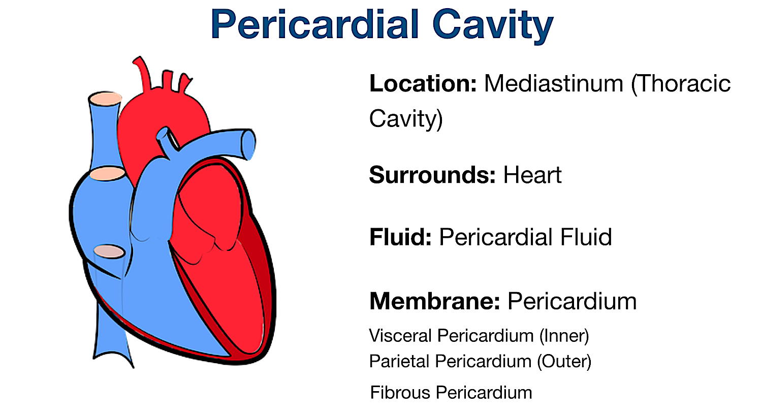
Pericardial Cavity Features
Location: Mediastinum (Middle Thoracic Cavity)
Surrounds: Heart, Roots of Great Vessels
Fluid: Pericardial Fluid
Membrane: Pericardium (Visceral, Parietal, and Fibrous Pericardium)
The pericardial cavity is located in the mediastinum within the thoracic cavity, and it surrounds the heart as well as the roots of the great vessels.
Similar to how the pleural cavity contained pleural fluid, the pericardial cavity contains a small amount of fluid called pericardial fluid.
Furthermore, similar to how the pleural cavity was lined by a double-layered serous membrane called the pleura, the pericardial cavity is lined by a double-layered serous membrane called the serous pericardium.
Just like the visceral and parietal layers of the lung, there is an inner visceral layer called the visceral pericardium and an outer parietal layer called the parietal pericardium.
The potential space between the visceral and parietal pericardium is the pericardial cavity.
There is also a fibrous pericardium that surrounds the outer serous parietal layer.
Let’s review all the layers in the next section to better understand them.
Pericardial Body Cavity: The pericardial cavity is located in the mediastinum and surrounds the heart. It contains pericardial fluid and is lined by the pericardium.
Pericardial Cavity Anatomy
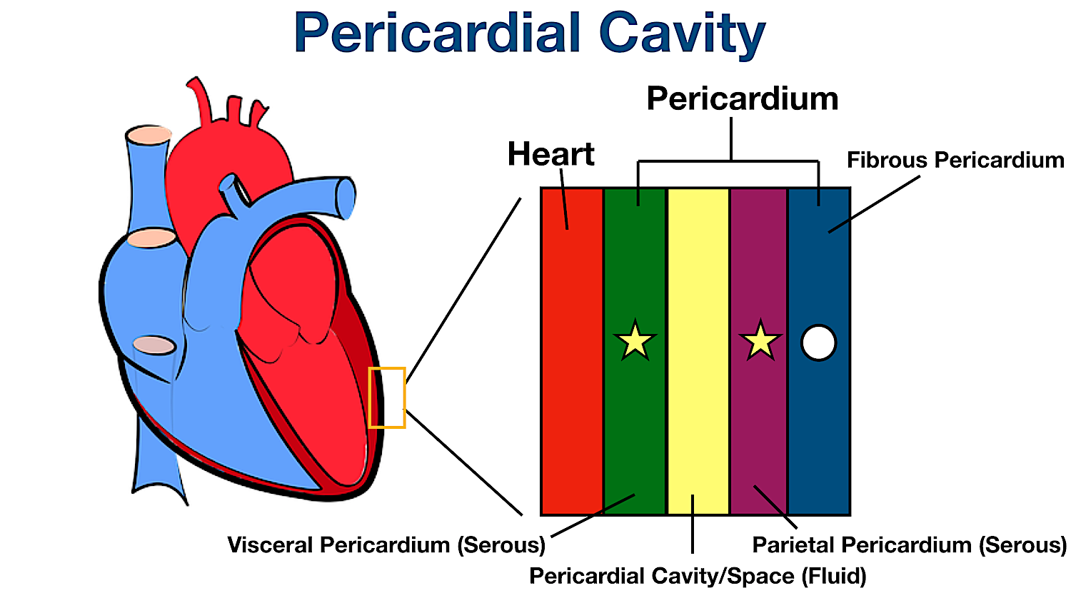
Let’s use the image below to better understand the features of the pericardial cavity and the layers of the pericardium.
If we zoom in on the outside of the heart, we can see how the heart is surrounded by the layers of the pericardium.
The pericardium is a sac that surrounds the heart and the roots of the great vessels.
The pericardium can be divided into:
A double-layered serous membrane (stars)
An outer fibrous membrane (circle)
The inner layer of the serous pericardium is the visceral pericardium, and it surrounds the heart.
This is similar to the visceral pleura that surrounded the lungs.
The outer layer of the serous pericardium is the parietal pericardium, and it lines the inside of the fibrous pericardium.
The potential space between the visceral and parietal serous pericardium is the pericardial cavity or space.
The pericardial cavity contains serous fluid called pericardial fluid that helps lubricate the membranes lining the cavity and heart to protect the heart as it beats.
The outermost layer of the pericardium is the fibrous pericardium, and it surrounds the serous pericardium.
In summary, the pericardial cavity in yellow surrounds the heart and is lined by the visceral and parietal serous layers of the pericardium.
Pericardial Cavity and Pericardium: The pericardium is the sac surrounding the heart. The pericardium can be subdivided into a double-layered serous pericardium (stars) and a fibrous pericardium (circle).
The visceral pericardium (green) is the inner serous layer that covers the heart (red). The parietal pericardium (purple) is the outer serous layer. The pericardial cavity (yellow) is the potential space between the visceral and parietal pericardial layers and contains pericardial fluid. The fibrous pericardium (blue) is the outermost layer of the pericardium.
Abdominopelvic Cavity
As mentioned above, the ventral cavity can be subdivided into 2 main parts that are separated by the diaphragm:
Thoracic Cavity
Abdominopelvic Cavity
If we go back to our flow chart, we now have a good understanding of the thoracic cavity.
The thoracic cavity can be subdivided into the right and left pleural cavities which surround the lungs, and the mediastinum which contains the heart, trachea, esophagus, great vessels, and thymus gland.
Within the mediastinum, there is another cavity called the pericardial cavity which surrounds the heart and the roots of the great vessels.
Now let’s move on to the abdominopelvic cavity.
Body Cavities Labeled Diagram: The ventral cavity can be subdivided into the thoracic cavity and the abdominopelvic cavity.
Abdominopelvic Cavity Features
Location: Inferior Ventral Cavity
Enclosed By: Rib Cage, Muscles, Vertebral Column, Diaphragm, Pelvis, Pelvic Floor
Houses: Abdominal and Pelvic Structures
Fluid: Peritoneal Fluid
Membrane: Peritoneum
The abdominopelvic cavity is the inferior portion of the ventral cavity located below the diaphragm, as highlighted in red below.
As the name suggests, the abdominopelvic cavity is in the abdomen and pelvis and consists of both the abdominal cavity and pelvic cavity.
Let’s discuss each of these cavities starting with the abdominal cavity followed by the pelvic cavity.
Abdominopelvic Body Cavity: Labeled diagram of the abdominopelvic cavity (red), which is the inferior portion of the ventral cavity below the diaphragm. It consists of the abdominal and pelvic cavities.
Abdominal Cavity
If we go back to our flow chart, we can subdivide the abdominopelvic cavity into 2 parts:
Abdominal Cavity
Pelvic Cavity
Let’s look at the abdominal cavity first followed by the pelvic cavity.
Body Cavities Labeled Diagram: The abdominopelvic cavity can be subdivided into the abdominal cavity and the pelvic cavity.
Abdominal Cavity Features
Location: Middle Ventral Cavity
Enclosed By: Rib Cage, Muscles, Vertebral Column, Diaphragm, Pelvic Inlet (Imaginary)
Houses: Liver, Stomach, Pancreas, Spleen, Kidneys, Intestines
Fluid: Peritoneal Fluid
Membrane: Peritoneum (Visceral Peritoneum and Parietal Peritoneum)
The abdominal cavity is the middle portion of the ventral cavity (or the superior portion of the abdominopelvic cavity) as highlighted in red below.
The abdominal cavity is enclosed mainly by the rib cage, abdominal muscles, and the vertebral column.
The diaphragm forms the superior boundary of the abdominal cavity, and an imaginary line at the pelvic inlet forms the inferior boundary between the abdominal and pelvic cavities.
Some of the main contents of the abdominal cavity include the liver, stomach, pancreas, spleen, kidneys, and intestines.
Similar to the pleura and pericardium, the abdominal cavity is lined with a double-layered serous membrane called the peritoneum.
The inner visceral peritoneum lines most of the internal abdominal organs or viscera, and the outer parietal peritoneum lines the walls of the abdominal cavity.
The peritoneal cavity is the potential space between the visceral and parietal peritoneal layers, and it contains a small amount of fluid called peritoneal fluid.
The peritoneal fluid helps lubricate the membranes that line the peritoneal cavity and most of the abdominal organs.
Abdominal Body Cavity: Labeled diagram of the abdominal cavity (red) and its features, including its main organs (liver, stomach, pancreas, spleen, kidneys, and intestines), membrane (peritoneum), and fluid (peritoneal fluid).
Abdominal Cavity Anatomy
The terms peritoneal cavity and abdominal cavity are different, so let’s take a closer look at these features.
Below is a side view of the small intestine in the abdominal cavity.
The small intestine is attached to the posterior abdominal wall by the mesentery.
The peritoneum is a continuous serous membrane that folds on itself to create 2 layers (similar to the pleura and serous pericardium).
The inner layer is called the visceral peritoneum and it covers most of the internal visceral organs of the abdominal cavity (shown in green).
The outer layer is called the parietal peritoneum and it lines the walls of the abdominal cavity (shown in red).
There is a potential space between the visceral and parietal layers of the peritoneum called the peritoneal cavity or space, and it contains serous fluid called peritoneal fluid that helps lubricate the peritoneum.
The peritoneal cavity is exaggerated in the image below since the other internal organs are removed.
In summary, the abdominal cavity is the entire space enclosed by the rib cage, abdominal muscles, vertebral column, diaphragm and pelvic inlet.
Whereas the peritoneal cavity is the potential space between the visceral and parietal peritoneum.
This becomes important because some structures are located in the abdominal cavity, but not “within” the peritoneum - more on that below!
Abdominal Body Cavity: Labeled diagram of the abdominal cavity showing the small intestine (brown) covered by the visceral peritoneum (inner green layer), and the parietal peritoneum (outer red layer) lining the abdominal cavity.
The peritoneal cavity is the potential space between the visceral and parietal peritoneum and contains peritoneal fluid.
Intraperitoneal Organs/Structures
Organs/structures that are covered by the visceral peritoneum are known as intraperitoneal organs.
Examples of intraperitoneal organs include the liver, stomach, spleen, jejunum, and ileum to name a few.
Intraperitoneal structures are “within” the peritoneum.
“Within” is in quotation marks as the organs are not technically inside the peritoneal cavity (the potential space between the visceral and parietal peritoneum).
Remember intraperitoneal organs are covered by visceral peritoneum, so they cannot technically be in the peritoneal space.
You might recall from the medical prefix lecture the prefix “intra-” means inside, internal, interior, inner, or within.
This can help you remember the meaning of intraperitoneal.
See image below as a reference.
Extraperitoneal Organs/Structures
There are also structures in the abdominal cavity that are extraperitoneal, meaning they are “outside” of the peritoneum.
More specifically, extraperitoneal structures are not covered by visceral peritoneum.
For example, structures could be retroperitoneal (behind the peritoneum) or subperitoneal (below the peritoneum) - see below!
You might remember from our medical prefix lecture the prefix “extra-” means outside, external, exterior, or outer.
This can help you remember the meaning of extraperitoneal.
Retroperitoneal Organs/Structures
Structures located behind the peritoneum or peritoneal cavity are called retroperitoneal structures.
They are called retroperitoneal structures because they are behind the peritoneum in the retroperitoneal space.
You may recall from our medical prefix lecture the prefix “retro-” means back, behind, or backward.
Retroperitoneal structures are covered by the parietal peritoneum anteriorly and attached to the posterior abdominal wall posteriorly.
See the image below illustrating a retroperitoneal structure with the star.
Example retroperitoneal organs located in the retroperitoneal space include the kidneys, adrenal glands, and part of the pancreas.
Subperitoneal Organs/Structures
Structures can also be located below or inferior to the peritoneum or peritoneal cavity.
They are called subperitoneal structures because they are below or inferior to the peritoneum or peritoneal cavity.
You might remember from our medical prefix lecture the prefix “sub-” means below, beneath, or under.
See the image below illustrating a subperitoneal structure with a circle.
An example subperitoneal organ is the bladder.
Again, make sure you know the difference between the abdominal cavity and peritoneal cavity because some structures may be in the abdominal cavity and not be intraperitoneal (not covered by visceral peritoneum).
The abdominal cavity is the entire cavity enclosed by the rib cage, abdominal muscles, vertebral column, diaphragm, and pelvic inlet.
Whereas the peritoneal cavity is the potential space between the visceral and parietal peritoneum.
Abdominal Body Cavity: Labeled diagram showing intraperitoneal organs (small intestine - brown) covered by visceral peritoneum, retroperitoneal organs (star) behind the parietal peritoneum, and subperitoneal organs (circle) below the parietal peritoneum.
Pelvic Cavity
As mentioned above, the abdominopelvic cavity can be subdivided into 2 parts:
Abdominal Cavity
Pelvic Cavity
We learned the abdominal cavity above.
Now let’s discuss the features of the pelvic cavity.
Body Cavities Labeled Diagram: The abdominopelvic cavity can be subdivided into the abdominal cavity and the pelvic cavity.
Pelvic Cavity Features
Location: Inferior Ventral Cavity
Enclosed By: Pelvis, Pelvic Floor Muscles, Pelvic Inlet (Imaginary)
Houses: Bladder, Reproductive Organs, Pelvic Portion of Colon, Rectum
Fluid: Peritoneal Fluid
Membrane: Peritoneum (Visceral Peritoneum and Parietal Peritoneum)
The pelvic cavity is the inferior portion of the ventral cavity.
It is enclosed by the pelvis and pelvic floor muscles.
The pelvic cavity is continuous with the abdominal cavity superiorly at the pelvic inlet.
The pelvic cavity mainly houses the urinary bladder, reproductive organs, pelvic portion of the colon, and rectum.
Since the pelvic cavity is continuous with the abdominal cavity, the fluid and membranes are the same.
The pelvic cavity is lined by the peritoneum which includes both the inner visceral peritoneum and outer parietal peritoneum just like we saw with the abdominal cavity.
The peritoneal cavity is the potential space between the visceral and parietal peritoneum, and it contains a small amount of peritoneal fluid.
Pelvic Body Cavity: Labeled diagram of the pelvic cavity (red) and its features, including the main organs (bladder, reproductive organs, pelvic portion of colon, and rectum), membrane (peritoneum), and fluid (peritoneal fluid).
Remember some of the structures in the pelvic cavity, such as the bladder, are subperitoneal (below the peritoneum) as illustrated by the blue circle below.
In summary, the pelvic cavity is the entire cavity enclosed by the pelvis and pelvic floor muscles (in green below), whereas the peritoneal cavity is the potential space between the visceral and parietal peritoneum.
Pelvic Body Cavity: Labeled diagram showing the pelvic cavity (green) and a subperitoneal structure in the pelvic cavity (blue circle) below the parietal peritoneum.
Body Cavity Flow Chart and Table
We now have a good understanding of the major body cavities by organizing them and breaking them down with the flow chart below.
Body Cavities Labeled Diagram: Summary flow chart of the major body cavities.
Here is a table that summarizes everything we learned to also make it easier for you.
Body Cavities Table: Summary of the major body cavities and features including their main organs, membranes, and fluid.


